Cyathus striatus
Scientific name: Cyathus striatus (Huds.) Willd.
Derivation of name: Cyath- means "cup." Striat-
means "finely furrowed" or
"lined" (striate) in reference
to the grooved inner
surface.
Synonyms: Peziza striata Huds.
Common name(s): Splash cups; Fluted bird's nest.
Phylum: Basidiomycota
Order: Agaricales
Family: Agaricaceae
Occurrence on wood substrate: Saprobic; clustered on
wood chips, bark, fallen branches; summer through fall.
Dimensions: Vase-shaped cups are up to 2 cm tall and
1cm wide.
Sterile nest surfaces: Exterior surface dark or grayish-
brown and covered with shaggy or wooly hairs; interior
surface distinctly grooved; shiny; pale gray or grayish-brown;
young vases are covered by a whitish membrane.
Fertile tissue: Gray peridioles ("eggs") occupy the bottom
of the vase-shaped cups; each egg is attached by a thread-
like
cord (funiculus) to the inner cup wall.
Comments:
When an egg is ejected, the trailing cord
(funiculus)
helps it adhere to an object it encounters.
More information at MushroomExpert.com:
More information at TomVolkFungi.net:
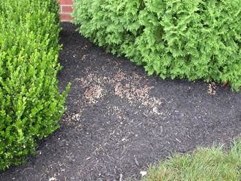
Figure 1. Landscape mulch is a great place to look for
bird's nest fungi. Photo © Gary Emberger.
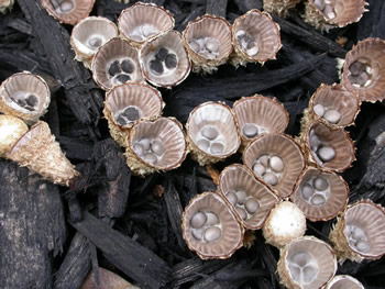
Figure 2. A closer view at some of the "nests" in Figure 1.
The eggs (peridioles) of Cyathus striatus are described as
gray or dark. Note in the specimens above that the eggs
are initially covered by a whitish membrane.
Photo © Gary Emberger.
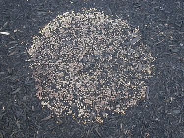
Figure 3.
This colony of bird's nest fruit bodies
is over two
feet in diameter. Assuming the colony started in the center,
the circular shape of the colony
reflects the uniform
nature of
the bark mulch substrate the
mycelium is digesting.
Photo © Gary Emberger.
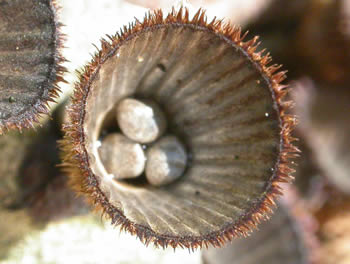
Figure 4. The hairy outside, grooved inside, and gray
peridioles make Cyathus striatus a distinctive fungus.
Photo © Gary Emberger.
%20Ringwood%20NJ%20Tom%20Bigelow.jpg)
Figure 5. This award-winning photograph of Cyathus
striatus features the specimens growing on a carpet of green
moss. Photo © Tom Bigelow.
%20Ringwood%20NJ%20Tom%20Bigelow.jpg)
Figure 6. A close-up of three specimens in Figure 5 highlights
the shaggy, wooly exterior of this species.
Photo © Tom Bigelow.
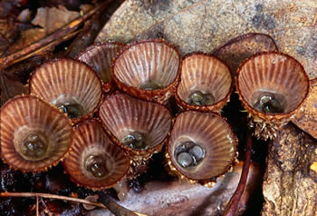
Figure 7. Forces resulting from a drop
of water striking
the
inside of the "splash" cup eject the eggs from the cup.
Photo © Pam Kaminski.
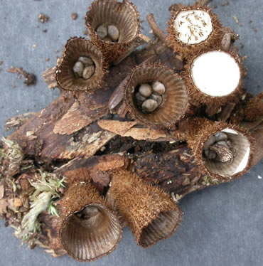
Figure 8. The nests on the right still have membranes or
remnants of membranes around the rim of the nest.
Photo © Gary Emberger.
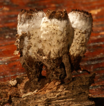
Figure 9. The whitish growths on these splash cups are the
mycelia of the ascomycete Trichoderm latizonatum
(synonym Hypocrea
latizonata), a parasite
only known to
occur on Cyathus striatus.
Photo © John Dawson.