Sphaerobolus stellatus
Scientific name: Sphaerobolus stellatus Tode
Derivation of name: Sphaer- means "sphere" and
bol- means "thrower."
Stell- means "star."
Synonyms: Carpobolus stellatus (Tode) Desm.
Common name(s): Cannon fungus; Sphere thrower;
Artillery fungus; Shotgun fungus.
Phylum: Basidiomycota
Order: Geastrales
Family: Geastraceae
Occurrence on wood substrate: Saprobic; solitary or
grouped on decaying wood (e.g., landscape bark mulch),
sawdust, herbaceous debris, or dung; spring through fall.
Dimensions: The fruit bodies are 1-3 mm wide.
Sterile nest surfaces: Yellow-orange, becoming whitish.
The nest opens by splitting into 4-9 starlike rays.
Fertile inner tissue: Single peridiole ("egg") about 1 mm
in diameter; dull yellow
to reddish-brown to dark brown.
Edibility: Are you kidding? Arora states that "Several
hundred would be needed for a mouthful!"
Comments:
The peridioles are sticky and will glue
themselves to objects such as house siding, windows,
and
cars. Shredded hardwood (tanbark) seems most prone
to supporting this fungus. To avoid the next-to-impossible-
to-get-off little tarry black
dots, use mulches in areas next
to houses and cars
that do not support the growth of this
fungus.
Recommended types are pine bark mulch or cedar
or cypress
mulch. Recent studies (Geml, J., et al., 2005)
indicate that three
species of Sphaerobolus are in the
Northeast, one of
which, Sphaerobolus ingoldii, was
determined to be new. The diameter of its peridioles is
about 1 mm.
The other two species, S.stellatus and S.
iowensis, have peridioles about 1.5 mm in diameter but can
otherwise only be differentiated from each other using
micromorphological features. As a result, many specimens
identified as S. stellatus are likely to be S. iowensis and
vice versa.
More information at TomVolkFungi.net:
More information at:
www.personal.psu.edu/faculty/d/d/ddd2/
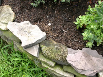
Figure 1.
Do you see the penny lying on the landscape
mulch? Photo © Gary Emberger.
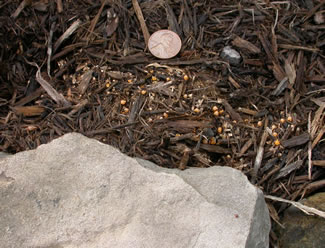
Figure 2.
Sphaerobolus stellatus is fruiting on the
mulch between the penny and the rock.
Photo © Gary Emberger.
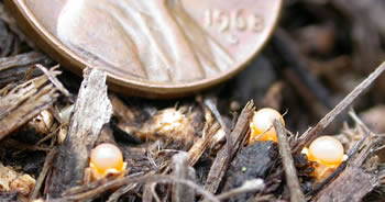
Figure 3. Several specimens of the fungus are growing in
the foreground. This
organism is tiny!
Photo © Gary Emberger.
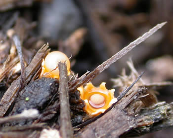
Figure 4. Note the star-shaped peridium and the single
egg
(peridiole) within. Photo © Gary Emberger.
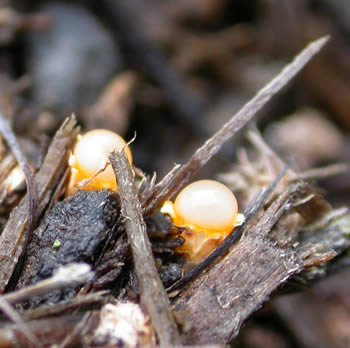
Figure 5. Here is the same specimen as in Figure 3. The
inner
layers have turned inside out, leaving behind a whitish
ball on
top of the rays. The peridiole was shot away.
Photo © Gary Emberger.
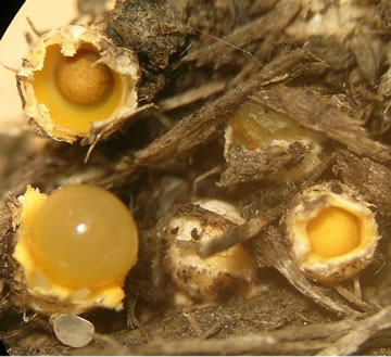
Figure 6. The peridium is just beginning to split apart in the
specimen on the far right, exposing the single peridiole.
Photo © John Dawson.
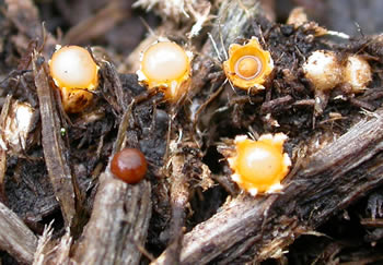
Figure 7. Several stages of development are in view here.
The
brown, shiny sphere to the lower left is a peridiole that,
for
whatever reason, did not make it very far. These "eggs"
are
capable of being propelled up to 6 m.
Photo © Gary Emberger.
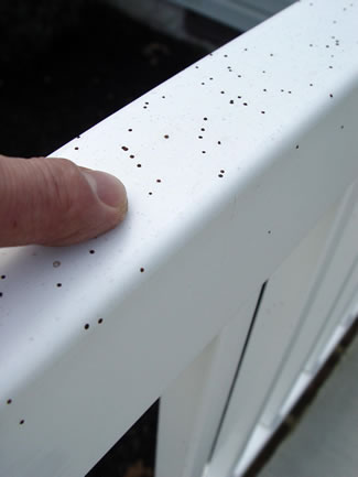
Figure 8.
The sticky projectiles of the artillery fungus
will glue themselves to whatever surfaces they strike.
Photo
© George Weigel.

Figure 9. Although each S. stellatus specimen shoots
only one peridiole, the combined output of a well-
populated bed of mulch can literally "pepper" surfaces
such as siding, downspouts, cars, doors, etc.
Photo © George Weigel.