Coprinellus micaceus
Scientific name: Coprinellus micaceus (Bull.) Vilgalys,
Hopple & Jacq. Johnson
Derivation of name: Copr- means "dung" and ellus is
diminutive. Micaceus means "glistening" and describes
the shiny particles on the cap.
Synonyms: Coprinus micaceus (Bulliard:Fries) Fries
Common name(s): Mica cap.
Phylum: Basidiomycota
Order: Agaricales
Family: Psathyrellaceae
Occurrence on wood substrate: Saprobic; densely clustered
around stumps, wood debris, at the base of standing dead or
dying trees or in grassy areas from buried wood; April through
October.
Dimensions: Caps 2-5 cm wide; stipes 2.5-8 cm tall and 2-5
mm thick.
Cap: Reddish-brown to tawny to ochre-brown, becoming
grayish particularly near the margin; surface
covered by
glistening granules that are soon lost; cap radially
lined almost
to the center.
Gills: Attached to nearly free; white, becoming black and inky
with age but not entirely dissolving.
Spore print: Black.
Stipe: White
Veil: Absent.
Comments: This species used to be in the genus Coprinus
but
DNA studies
radically revised the taxonomy of that
genus: Coprinus was retained for a small number of species,
several new genera were created, and the members of the
family Coprinaceae were split between two families -
Psathyrellaceae and Agaricaceae.
More information at TomVolkFungi.net:
More information at MushroomExpert.com:
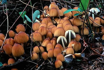
Figure 1. A clump of mica cap. Photo © Pam Kaminski.
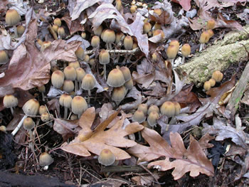
Figure 2. Older specimens become grayish. The
woody
debris these specimens are growing on is mostly obscured
by the fallen leaves. Photo © Gary Emberger.
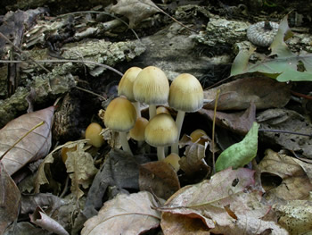
Figure 3. This clump of mica caps was growing on a log in
a wood pile but the fallen leaves obscured the connection
of the mushrooms to the wood. Photo © Gary Emberger.
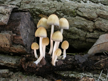
Figure 4. The clump of mica caps in Figure 2 with the
leaves removed. Not only is the substrate revealed
but also the elongation of the stems to position the caps
above the leaves. Photo © Gary Emberger.
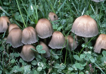
Figure 5. The caps are conspicuously striate. Photo ©
William Roody.
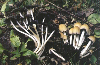
Figure 6. The gills of the mature specimens on the left are
partially auto-digested into an inky fluid. Photo © Larry
Grand.
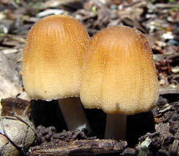
Figure 7. The glistening mica-like particles are visible on
these young specimens. Photo © David Work.
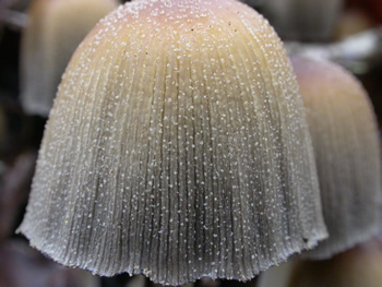
Figure 8. Mica-like granules on an older specimen.
Photo © Gary Emberger.
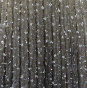
Figure 9. The mica-like granules on the cap surface are the
remnants of a
universal veil. Photo © Gary Emberger.
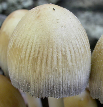
Figure 10. The mica-like particles quickly fall away with
age
or may be washed off by rain. They are often not very
evident on older
specimens. Photo © Gary Emberger.