Galerina marginata
Scientific name: Galerina marginata (Batsch) Kühner
Derivation of name: Galer- refers to a "helmut or fur cap"
and ina is the diminutive form. Marginata refers to martin,
edge,
or border; autumnalis (of the older name) implies
being
found during
the fall.
Synonyms: Galerina autumnalis (Peck) A.H. Sm. &
Singer, Pholiota autumnalis Peck
Common name(s): Deadly Galerina.
Phylum: Basidiomycota
Order: Agaricales
Family: Hymenogastraceae
Occurrence on wood substrate: Saprobic; in small groups or
clusters on decaying deciduous and conifer wood such as logs,
stumps, and buried wood; May through June, October through
November.
Dimensions: Caps 2.5-6.5 cm wide; stipes 2.5-10 cm long
and 0.3-1 cm thick.
Cap: Sticky to dry, smooth, yellow-brown to dark brown.
Gills: Attached; yellowish, becoming rust colored.
Spore print: Rusty brown.
Stipe: Whitish above, browish toward base; whitish mycelium
at point of attachment.
Veil: Membranous, white, evanescent. Appearing brown from
spore deposit. Due to the evanescent nature of the ring,
there may only be a brownish ring zone on the stipe or, in
some cases, no evidence of a partial viel at all.
Comments: The common name refers to the deadly
phallotoxins and amatoxins found within this species. This
mushroom is widely known by its synonym, G. autumnalis.
.
More information at MushroomExpert.com:
More information at TomVolkFungi.net:
.jpg)
Figure 1.
Fruiting of Galerina marginata on a rotting log.
Photo © George Morrison.
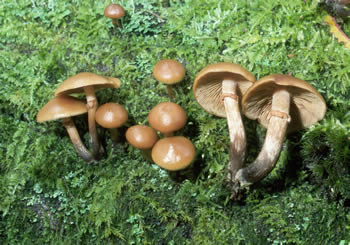
Figure 2. Deadly galerina on a moss-covered log. Note
the veils, overall brownish color, and shiny (sticky) cap.
Photo © William Roody.
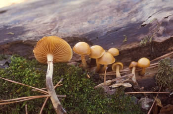
Figure 3. Mushroomers often ignore "little brown
mushrooms" due to their reputation as being difficult to
identify. Because of its deadly toxins, there is reason to
have a good mental image of this particular little brown
mushroom. Photo © Larry Grand.
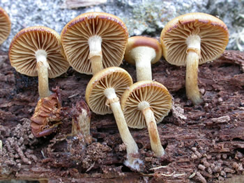
Figure 4. Note the partial veil covering the gills of the
youngest specimen. A whitish mycelium is evident at
the base of the stipe of the specimen in the foreground.
Photo © Gary Emberger.
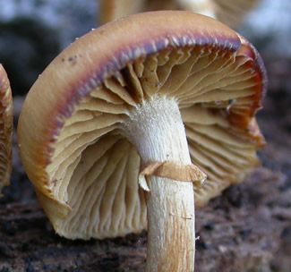
Figure 5. A distinctly membranous partial veil.
Compare to Figure 6.
Photo © Gary Emberger..
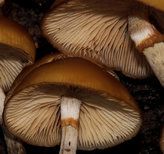
Figure 6. The collapsed
or deteriorated partial veils of
these
specimens are
very different in appearance from
the veil
pictured in Figure 5. Photo © Al Simpson.