Pluteus cervinus
Scientific name: Pluteus cervinus (Schaeff.) P. Kumm.
Derivation of name: In reference to this genus of mushrooms,
the etymology of Pluteus is difficult to determine. Cervin-
pertains to "deer" or "fawn-colored" but according to the
website authors below, the reference to deer is not to their
color but rather to the antler-like (horn-like)
projections at the
tips of the pleurocystidia - sterile cells covering
the gill
surfaces (Figure 5).
Synonyms: Pluteus atricapillus (Batsch) Fayod
Common name(s): Fawn mushroom; Deer mushroom.
Phylum: Basidiomycota
Order: Agaricales
Family: Pluteaceae
Occurrence on wood substrate: Saprobic; solitary or in
small groups on or around decaying deciduous, and less
often, conifer stumps and logs.
May through October.
Dimensions: Caps 3-12 cm wide; stipes 5-10 cm long and
0.5-1 cm thick.
Cap: Smooth, sometimes streaked with radially oriented fibers;
variable in color: brown to grayish-brown to pale cinnamon-
brown.
Gills: Free; white at first, becoming salmon-pink.
Spore print: Salmon-pink.
Stipe: White to grayish-brown.
Veil: Absent.
Comments: Recent DNA studies suggest a number of
cryptic (look-alike) species exist under the P. cervinus
name.
Given this reality, some simply recognize a
Pluteus cervinus "group."
Consult the MushroomExpert
link below for information to help distinguish among
some of these cryptic species such as P. petasatus.
For example, P. cervinus is typically a woodland species
whereas P. petasatus (Figures 6, 7) is often found in urban
settings, growing on dead deciduous wood but also on
wood chips and sawdust piles. Depending on the specimen,
microscopic examination may be necessary to confirm
identity.
More information at MushroomExpert.com:
More information at TomVolkFungi.net:
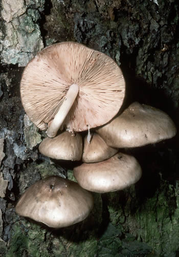
Figure 1. Fawn mushroom fruiting on wood.
Photo © William Roody.
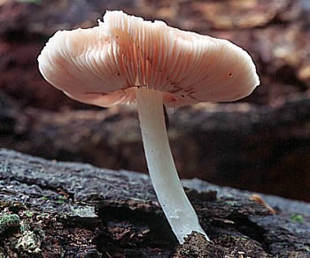
Figure 2. A beautiful view of the pink gills and white
stipe of
Pluteus cervinus. Photo © Pam Kaminski.
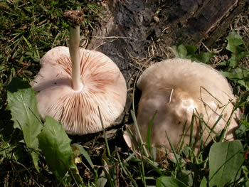
Figure 3.
The gills are initally white but become pinkish
as the pinkish spores mature. Photo © Gary Emberger.
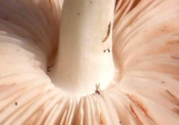
Figure 4. The distinctly free gills of Pluteus cervinus.
Photo © Gary Emberger.
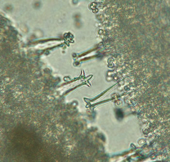
Figure 5. Microscope preparation of Pluteus cervinus
gill tissue. The long, projecting, hollow-looking cells are
pleurocystidia, sterile cells which cover the gill surfaces.
The horn-like (antler-like) projections at the tips of the
pleurocystidia help to characterize this species. This
picture of "horned" pleurocystidia
is one of the very few
microscopic traits pictured on this
website. While the use
of a microscope to find these cystidia is not necessary to
identify this species, the picture does illustrate the wealth
of additional information at the microscopic level which
can be
invaluable when working out mushroom
identifications. Photo © Gary Emberger.
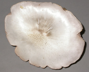
Figure 6. Pluteus petasatus is one of the cryptic
companions of P. cervinus. It is common on wood chips
and woody mulch whereas
Pluteus cervinus is typically
a woodland species. It's cap coloration is also typically
lighter than that of
Pluteus cervinus.
Photo © Gary Emberger.
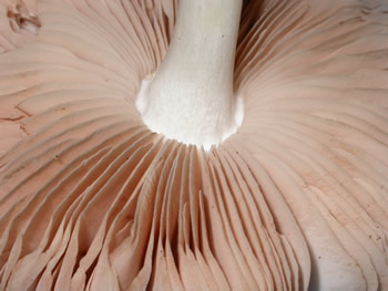
Figure 7. The free, salmon-pink colored gills of P.
petasatus. These features are typical of all Pluteus spp.
Photo © Gary Emberger.