Trichaptum abietinum
Scientific name: Trichaptum abietinum (Dicks.) Ryvarden
Derivation of name: Trichaptum means "with clinging
hairs"; abietinum means "inhabiting the fir tree" in reference
to firs (Abies), one of its common conifer substrates.
Synonymy: Polyporus abietinus Dicks.: Fr.;
Hirschioporus abietinus (Dicks. ex Fr.) Donk
Common names: Violet-toothed polypore.
Phylum: Basidiomycota
Order: Polyporales
Family: Polyporaceae
Occurrence on wood substrate: Saprobic; solitary or in
overlapping clusters, sometimes laterally fused, on dead
conifer wood, rarely on deciduous wood; year-round.
Dimensions: Caps 1-4 (rarely more than 1 cm) cm wide
and 1-2 mm thick.
Upper surface: Whitish to grayish or darker; somewhat
zonate; greenish if covered with algae; margin with violet
coloration.
Pore surface: Bright purplish at first, fading to ochraceous
with violet tones restricted to the margin; pores 2-4 per mm,
becoming tooth-like.
Comments: Compare to Trichaptum biforme which occurs
almost always on hardwoods and is generally larger. Because
there
are two other
Trichaptum species (not illustrated)
which have
purplish lower surfaces at
least when young and
which grow
on conifers, some care is warranted before
too
quickly
labeling a given
specimen as T. abietinum. One of
these is
Trichaptum laricinum which
has
a
gill-like lower
surface. The other is Trichaptum fuscoviolaceum which
has a lower surface described as bearing short gill-like
sections which become tooth-like, resulting in lines or rows
of teeth.
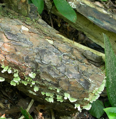
Figure 1. Trichaptum abietinum on conifer log.
Photo © Gary Emberger.
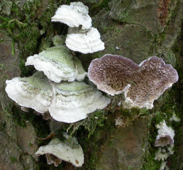
Figure 2. The specimen on the right is turned upside down
to show
the violet-colored pore surface.
Photo © Gary Emberger.
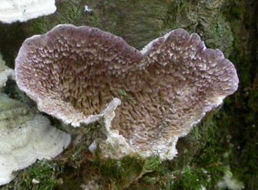
Figure 3. A close-up of the specimen in Figure 2. When
young, Trichaptum abietinum has 4-6 pores per mm. At
maturity, the walls forming the pores become thin and deeply
lacerate
(i.e., torn, toothed) so that the entire lower surface
looks tooth-like. Photo © Gary Emberger.
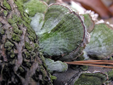
Figure 4. Trichaptum abietinum on conifer wood. Note the
purple marginal band. Photo © Dianna Smith.
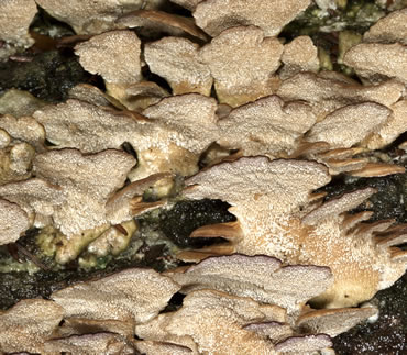
Figure 5. The violet coloration in this specimen is restricted to
a narrow band along the margin of the tooth-like pore
surface. Photo © Dianna Smith.
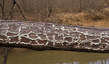
Figure 6. The tiny shelves of Trichaptum abietinum are
located between the large bark plates of this conifer log.
The fungus is growing specifically on the sapwood.
Photo © Larry Grand.
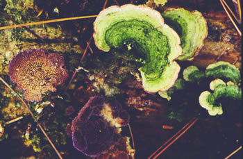
Figure 7. Specimens of Trichaptum abietinum often
develop a conspicuous growth of green algae on the cap.
Photo © John Plischke III.