Phallus ravenelii
Scientific name: Phallus ravenelii Berk. & M. A. Curtis
Derivation of name: Phallus means "male member,
penis." Ravenelii is named in honor of Henry William
Ravenel (1814-1887) a South Carolina mycologist and
botanist.
Synonyms: Aedycia ravenelii (Berk. & M. A. Curtis)
Kuntze
Common name(s): Ravenel's stinkhorn.
Phylum: Basidiomycota
Order: Phallales
Family: Phallaceae
Occurrence on wood substrate: Saprobic on lignin-rich
humus in gardens, flowerbeds, and woods; solitary or
clustered on wood debris, wood chips, sawdust, decaying
stumps and logs in the woods; August through October.
Dimensions: Fruit body up to 20 cm tall; head up to 4 cm
wide; stalk up to 3 cm thick.
Description: Fruit body at first a white to pinkish-lilac egg-
like stage, resembling a puffball. The "egg" is attached to the
substrate by white to pinkish mycelial strands (rhizomorphs).
The outer wall (peridium) of the egg splits and a hollow,
spongy, whitish stalk expands bearing a head covered with a
slimy, olive-green fetid spore mass. Under the slimy spore
mass, the head is smooth or granular to somewhat wrinkled
but is not deeply pitted and ridged.
Comments: Flies are attracted to the fetid, slimy mass
and serve to disperse the spores. Phallus impudicus is very
similar in overall appearance and habitat but
the head is
deeply pitted and ridged.
For Phallus ravenelii:
More information at MushroomExpert.com:
For Phallus impudicus:
More information at MushroomExpert.com:
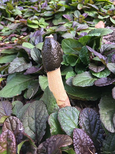
Figure 1. Phallus ravenelii growing in a garden bed. It was
definitely not planted by the owner.
Photo © Melissa Kurtz.
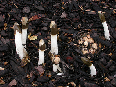
Figure 2. Cluster of mature specimens and a cluster of "eggs."
Photo © Gary Emberger.
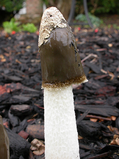
Figure 3.
The spongy stipe expands rapidly. In this specimen,
a portion of the peridium was carried aloft - like a skullcap!
Photo © Gary Emberger.
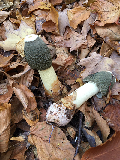
Figure 4.
The stinkhorn in the foreground was collected and
placed on its side. The slightly
pinkish peridium of the "egg"
and the pinkish
rhizomorphs
atached to it are to the left. The
slime-covered head is at the
right.
Photo © Gary Emberger
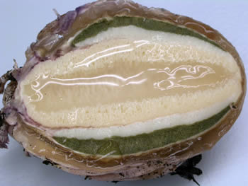
Figure 5. A bisected egg of Phallus ravenelii showing the
outer pinkish-lilac peridium, the unexpanded stipe filled
with a gelatinous material in the center and the dark
olive-green spore mass associated with the skirtlike head.
Photo © Gary Emberger.
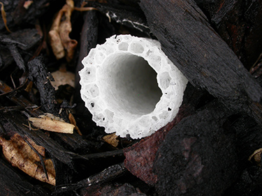
Figure 6. A cross section of the hollow stipe showing the
sponge-like wall of the stipe.
Photo © Gary Emberger.
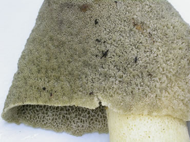
Figure 7. The lower margin of the skirtlike head is free from
the stalk. The outer surface of the head may be granular or
wrinkled but is not deeply pitted and ridged.
Photo © Gary Emberger.
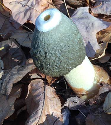
Figure
8. The head is attached to a white, mouthlike circlet
at the apex of the stipe. The hole surrounded by the
circlet tissue is continuous with the hollow stipe.
Photo © Gary Emberger.
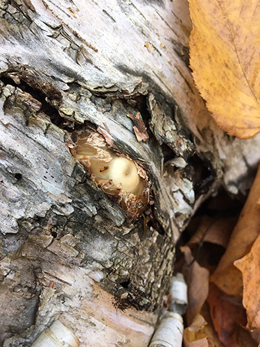
Figure
9. Phallus ravenelii emerging from its "egg"
located just below the bark of a birch tree.
Photo © Gary Emberger.
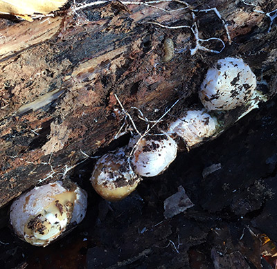
Figure 10. The loose, rotting bark of the birch tree in Figure 9
was lifted away, revealing a number of "eggs" and rhizomorphs
of
Phallus ravenelii.
Photo © Gary Emberger.
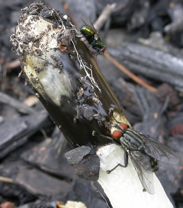
Figure 11. As with all stinkhorns, flies are attracted to
and consume the smelly spore mass.
Photo © Gary Emberger.
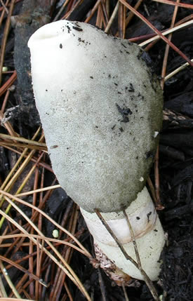
Figure 12. The appearance of the head after the
flies finish feasting. Photo © Gary Emberger.
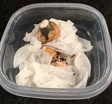
Figure 13. If mature stinkhorn "eggs" are placed in moist
paper toweling, they may continue along their
developmental pathway. This photo was taken at 4:50pm.
Photo © Gary Emberger.
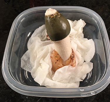
Figure 14. The specimen in Figure 13 at 10:30pm.
Photo © Gary Emberger.
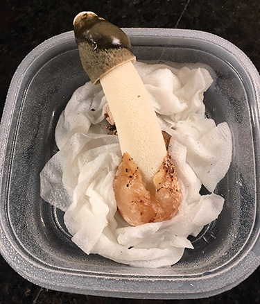
Figure
15. The specimen in Figure 14, photographed at
12:30pm the following day. The tray was kept outside and
flies have cleaned off quite a bit of the spore slime mass.
Photo © Gary Emberger.