Climacodon septentrionalis
Scientific name: Climacodon septentrionalis (Fr.)
P. Karst.
Derivation of name: Septentrional- means "northern."
Synonyms: Steccherinum septentrionale (Fr.) Banker;
Hydnum septentrionale Fr.
Common name(s): Northern tooth.
Phylum: Basidiomycota
Order: Polyporales
Family: Phanerochaetaceae
Occurrence on wood substrate: Parasitic; in dense
overlapping clusters on trunks of living deciduous trees,
particularly maple (Acer) and beech (Fagus); July through
October.
Dimensions: Individual caps up to 30 cm wide and from
2.5-5 cm thick at the base. Overlapping clusters of shelving
caps may be up to 80 cm high.
Description: Upper cap surfaces are whitish to creamy
yellow when young and become yellow-brown in age. Cap
surfaces are hairy to rough. Odor and taste when young are
not distinctive but the odor of old specimens is described as
like old, spoiled ham and the taste becomes bitter. The
crowded, whitish spines on the underside of the caps are 0.5-
2 cm long and have lacerated or ragged tips. Like the cap
surfaces, the spines become yellowish in age.
Edibility: Not considered edible.
Comments: This fungus looks like a polypore until the
spines
are noticed. It causes a heart rot of trees in urban
areas,
parks, and in forests.
More information at MushroomExpert.com:
More information at TomVolkFungi.net

Figure 1. Climacodon septentrionalis on a dying hardwood
tree at Mine Falls Park in Nashua, NH.
Photo © Michael Emberger.
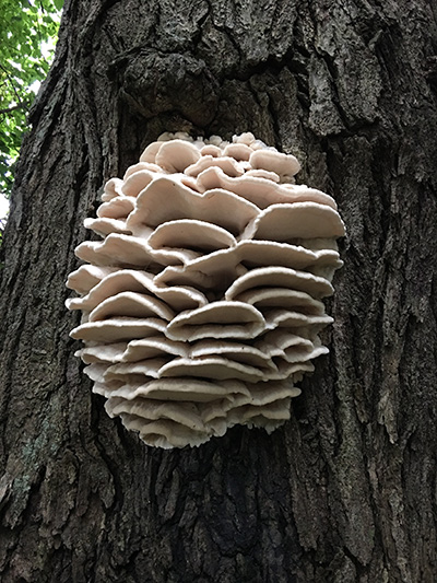
Figure 2. Close-up of the fungus in Figure 1. The overlapping
shelves often form a very symmetrical overall shape.
Photo © Michael Emberger.
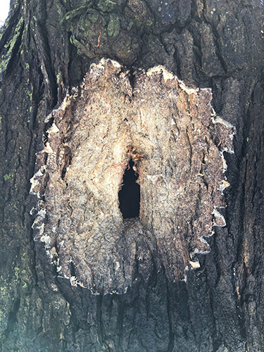
Figure 3.
Climacodon septentrionalis often grows from a
wound on the host tree. After the specimen in Figure 2
decayed and fell away, a large hole in the trunk was
revealed.
Photo © Michael Emberger.
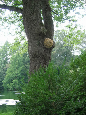
Figure 4. Specimen of Climacodon septentrionalis
growing on red maple (Acer rubrum) at Longwood
Gardens in Pennsylvania. Photo ©
Gary Emberger.
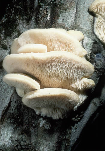
Figure 5. Climacodon septentrionalis looks like a
polypore until you see the spines under the caps.
Photo © William Roody.
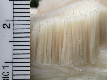
Figure 6. A portion of a cap was flipped over to examine
the
spines which are about 1 cm in length.
Photo ©
Gary Emberger.
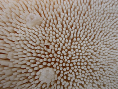
Figure 7. Tips of the teeth (spines). Photo © Gary Emberger.
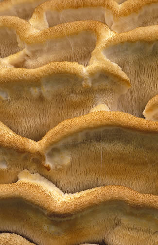
Figure 8. Older specimen of northern tooth with
yellow-brown discoloration. Photo © Fred Habegger.
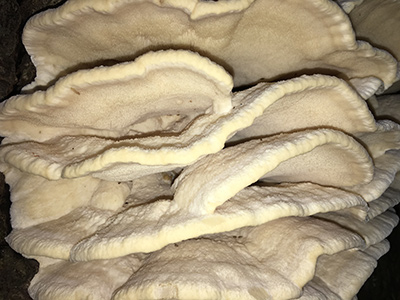
Figure 9. The upper cap surfaces of young specimens are whitish
to yellowish and hairy to rough
in texture.
Photo © Michael Emberger.
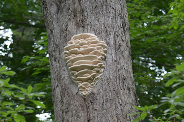
Figure 10.
Climacodon septentrionalis associated with a
crack on a maple (a different tree than the one in Figure 4)
at Longwood Gardens.
The overlapping caps progressively
decrease in size toward the top and bottom of the
cluster.
Photo © Don Davis.
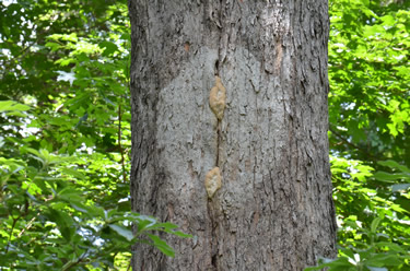
Figure 11.
This is the same tree as pictured in Figure 10. The
fruit body
either fell off or was knocked off or removed in
some manner but
the
fungus is re-emerging from the crack
in the trunk.
Photo © Don Davis.
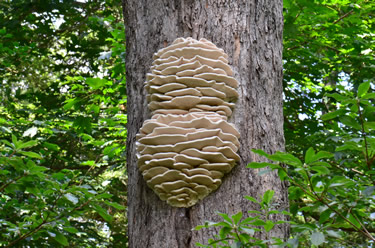
Figure 12. This photo was taken 1 month after the image in
Figure 11. The re-emerged fungus fruit body is even larger
than the original structure in Figure 10. Photo © Don Davis.
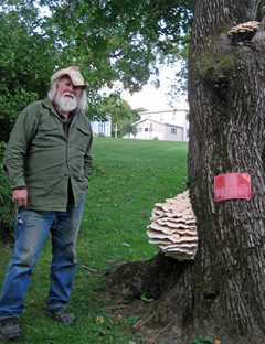
Figure 13. Northern tooth is often reported
high up on wounds of infected trees but this
low-to-the-ground specimen on a living Norway
maple (Acer platanoides) shows there are
often exceptions. Photo © Lynne Jones.