Pholiota squarrosa
Scientific name: Pholiota squarrosa (Vahl) P. Kumm.
Derivation of name: Phol- means "scale" referring to the
scaly caps of many species in the genus. Squarros-
means "with upright
scales,
rough, scurfy" (squarrose) in
reference to the
prominent scales.
Synonym: Agaricus squarrosus Vahl
Common name(s): Scaly Pholiota.
Phylum: Basidiomycota
Order: Agaricales
Family: Strophariaceae
Occurrence on wood substrate: Saprobic; typically in
cespitose clusters
on living or dead deciduous or conifer
wood,
sometimes at the
base of the tree; July through
November.
Dimensions: Caps 2.5-10 cm; stipes 5-10 cm long and
3-15
mm thick.
Cap: Dry; yellow-brown surface covered with pinkish-tan
or
brownish scales.
Gills: Attached; yellow at first, developing green tones,
rust-
brown at maturity.
Spore print: Brown.
Stipe: Dry; scaly like the cap.
Veil: Yellowish partial veil leaving persistent membranous
ring
or sometimes ring zone on upper stalk.
Edibility: Poisonous. Although some people eat it, it is
not
recommended as there are reports of gastric upset
following
ingestion.
Comments: Some specimens have an odor of onions or
garlic. Michael Kuo makes the point in his website
below that there is usually enough uncertainty due to age,
variable and overlapping traits, and weather conditions,
that microscopic analysis may be required to confirm
identifications for this and most Pholiota species.
More information at MushroomExpert.com:
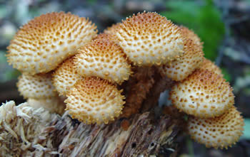
Figure 1. Typical cespitose cluster on wood. Photo©
David
Work.
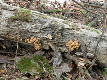
Figure 2. Clusters of Pholiota squarrosa on a log.
Photo © Gary Emberger.
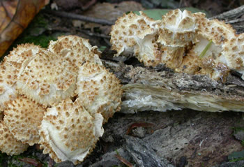
Figure 3. Clusters of Pholiota
squarrosa on the same log
shown in Figure 2. The outer cap
tissue was cracked and
peeling on most of the
mushrooms on this log. Our club
members attributed this to the hot and dry conditions
experienced by these developing mushrooms. Photo ©
Gary Emberger.
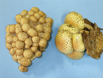
Figure 4. These specimens were collected and identified
during the 2003 NEMF foray. Photo © Gary Emberger.
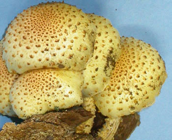
Figure 5. A closer
view of the specimens on the right in
Figure 4. Photo © Gary Emberger.