Poronidulus conchifer
Scientific name: Poronidulus
conchifer (Schwein.)
Murrill
Derivation of name: conchifer means "shell bearing."
Synonymy: Polyporus conchifer Schw.: Fr.; Trametes
conchifer (Schwein.) Pilat
Common names: Little nest polypore.
Phylum: Basidiomycota
Order: Polyporales
Family: Polyporaceae
Occurrence on wood substrate: Saprobic; in groups on
decaying deciduous wood; June through December.
Dimensions: Cups 0.5-2 cm wide; caps 1-5 cm wide.
Upper surface: Cups concentrically zoned, white and brown;
caps zoned with bands of white and grayish-white and pale
brown; radially wrinkled.
Pore surface: White to yellowish; pores 2-4 per mm.
Edibility: Inedible.
Comments: The cups may be found by themselves. They are
sterile and may be mistaken for a cup fungus or a bird's nest
fungus. If just the shelves are present, this fungus is difficult to
identify. The combination is distinctive.
More information at MushroomExpert.com
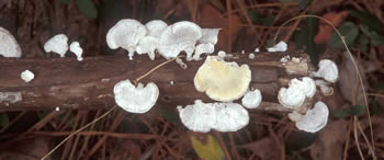
Figure 1. Trametes conchifer on wood. The cups are not
prominent in this photo. Note the yellowish pore surface of
the flipped-over specimen. Photo © Larry Grand.
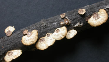
Figure 2. Various combinations of cups and shelf-like caps are
visible in these specimens. Photo © Gary Emberger.
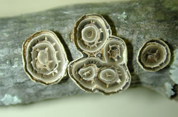
Figure 3. Sterile cups often form first and are typically
concentrically zoned. Photo © Tom Volk.
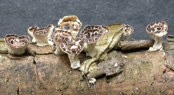
Figure 4. These cups are stalked. It's no wonder they are
mistaken for cup fungi. The cups function as splash cups
dispersing
asexual propagules called oidia.
Photo © Gary Emberger.
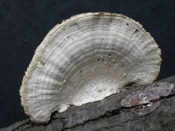
Figure
5. This cap shows no evidence of a cup. Note the
faint zonation and radial wrinkles. Photo © Gary Emberger.
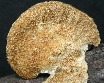
Figure 6.
The underside of the shelf-like cap reveals
the
pores of the fertile portion of the fungus. Photo © Gary
Emberger.
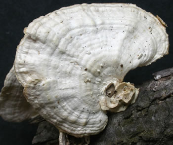
Figure 7. Just a little of the cup structure remains at the base
of the shelf. It's enough to identify the species with certainty.
Photo © Gary Emberger.
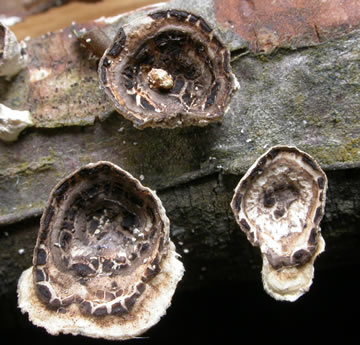
Figure 8. Shelves are beginning to develop from these cups.
Photo © Gary Emberger.
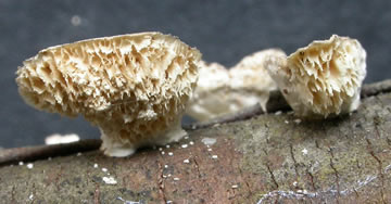
Figure 9.
Pores
can be seen on the underside of the cups of
Figure 8. Photo © Gary Emberger.