Trichaptum biforme
Scientific name: Trichaptum biforme (Fr.)
Ryvarden
Derivation of name: Trichaptum means "with clinging
hairs"; biforme means "with two forms or stages" in reference
to the pore surface which can be either poroid or toothlike.
Synonymy: Polyporus biformis Fr. in Kl., Polyporus
pergamenus Fr.; Trichaptum pargamenum (Fr.) G. Cunn.;
Hirschioporus pergamenus (Fr.) Bondartsev & Singer
Common names: Violet-toothed polypore.
Phylum: Basidiomycota
Order: Polyporales
Family: Polyporaceae
Occurrence on wood substrate: Saprobic; solitary to
overlapping clusters on dead deciduous wood, rarely on
conifers; year-round.
Dimensions: Caps 1-7.5 cm wide and up to 3 mm thick.
Upper surface: White to grayish or brownish, greenish if
covered by algae; margin often purplish; zonate; hairy.
Pore surface: Purplish at first, fading to buff or brownish but
usually retaining violet tints near margin; poroid at first with
pores 2-5 per mm, becoming toothlike in age.
Edibility: Inedible.
Comments: A very colorful polypore when young. It can
occur in great numbers on the substrate. Compare to
Trichaptum
abietinum which occurs almost always
on conifer
wood and is generally
smaller. Trichaptum
subchartaceum (not illustrated) is a boreal species that
grows only on Populus spp. (aspen, poplar) and does
not develop a conspicuous tooth-like lower surface. Be
sure to look at Figures 11 and 12 to become aware of
Phaeocalicium polyporaeum, a tiny saprobic ascomycete
often found on old specimens of T. biforme. A related
species,
Phaeocalicium curtisii grows on twigs and
branches of staghorn sumac (Rhus typhina).
More information at MushroomExpert.com:
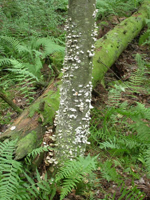
Figure 1. This birch (Betula) tree is covered with
Trichaptum biforme fruit bodies.
Photo © Gary Emberger.
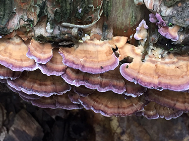
Figure 2. Purplish margins of young specimens.
Photo © Dorothy Smullen.
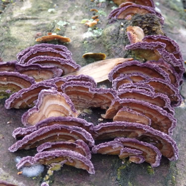
Figure 3. Zonate caps of young specimens of violet-toothed
polypore with intense
coloration along the cap
margins.
Photo © John Dawson.
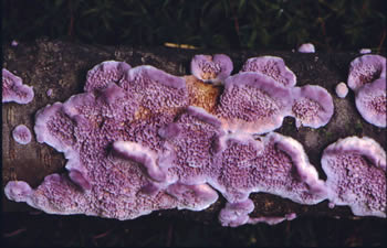
Figure 4.
Very young specimens may have vivid
violet coloration as evidenced here.
Photo © John Plischke III.
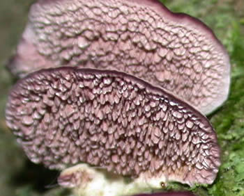
Figure 5. The tooth-like pore surface is quite evident
as
is the overall violet coloration. Photo © Tom Volk.
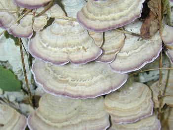
Figure 6. The violet coloration of Trichaptum biforme
fades over time. Here, the color is restricted to just
the
cap margins.
Photo © Tom Volk.
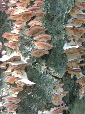
Figure 7.
The violet coloration
persists longest on
the tooth-like pore
surface. Even then, as observed
here, the color may be evident only at the pore
surface margins. Photo © Stephanie Depew.
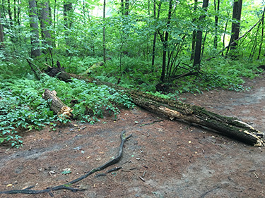
Figure 8. Trichaptum biforme is one of the most common
fungi in a hardwood forest, encountered on virtually every
foray. This fallen red maple tree is covered with old fruit
bodies of the fungus. Photo © Michael Emberger.
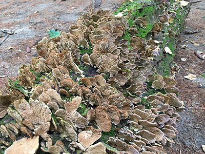
Figure 9. Fertile surfaces of Trichaptum biforme on the log
pictured in Figure 8. Old specimens like these are typically
devoid of color. Photo © Michael Emberger.
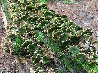
Figure 10.
The upper surfaces of old fruit bodies such as
these growing on the log pictured in Figure 8 are often green
due to the growth of algae.
Photo © Michael Emberger.
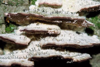
Figure 11. Tiny black club-like fungi are often present
on the
upper surface of
older Trichaptum biforme
fruitbodies.
The little clubs are Phaeocalicium
polyporaeum,
an ascomycete. They are sometimes
called fairy pins.
Photo © John Plischke III.
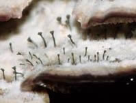
Figure 12. A closer view of Phaeocalicium polyporaeum,
a saprotroph on senescing tissue of of old Trichaptum
biforme specimens.
Photo © John Plischke III.