Radulomyces copelandii
Scientific name: Radulomyces copelandii (Pat.)
Hjortstam
&
Spooner
Derivation of name: TBD
Synonyms: Radulodon copelandii (Pat.) N. Maek.;
Hydnum copelandii Pat.
Common name(s): Asian beauty.
Phylum: Basidiomycota
Order: Agaricales
Family: Pterulaceae
Occurrence on wood substrate: Saprotroph; on
hardwood logs and dead standing trees, especially on
oak (Quercus)
and maple (Acer); year-round.
Dimensions: Individual fruit bodies up to 30 cm in length.
The spines can be quite long - up to 1.2 (1.4) cm.
Description: Resupinate fruit bodies develop in the cracks,
interstices, and
furrows of the bark of dead trees. The
densely crowded
spines are white to pale
yellowish, turning
buff to brownish in
age.
Edibility: Inedible.
Comments: In 2011, J. Ginns and Lawrence Millman
reported the
first known
occurrence of this species in the
Western
Hemisphere - in Massachusetts. It has since been
reported from Connecticut and Rhode Island. As far as I
am aware, the photographs on this page document the first
known report from Pennsylvania.
A 2018 report
documents the occurrence of a new Raduomyces
species in North America: R. paumanokensis J. Hormon,
B. Ortez, and K. Nakasone. It is similar to but different
than R. copelandii in appearance
and is pictured in
Figures 11 and 12 as a potential look-alike species.
More information at MushroomExpert.com
More information at Fungi.org:
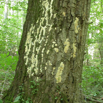
Figure 1. A fungus-festooned dead chestnut oak (Quercus
montana)
at Middle Creek Wildlife Management Area in
Pennsylvania in August, 2013. The species that caught my
attention
was the toothed fungus on the right-hand side of
the tree.
Photo © Gary Emberger.
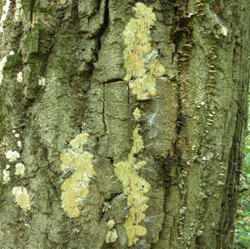
Figure 2. Fruit bodies of Radulomyces copelandii
develop in
the cracks and furrows of the bark.
Photo © Gary Emberger.
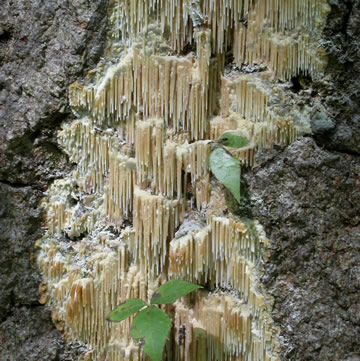
Figure 3. There are no stems or caps, the fruit bodies are
completely resupinate. Photo © Gary Emberger.
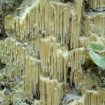
Figure 4. Lawrence Millman reports that Asain beauty fruits
year-round and that the spines are more likely to go from
whitish to brownish directly in the summer and fall. In the
winter, however, there is usually an intermediate yellowish
phase. Photo © Gary Emberger.

Figure 5 The slender, pointed spines are up to 1.4 cm long
and are round in cross section. Although microscopic
examination of the spores was helpful in confirming the
identification,
a positive field
identification is possible based
on the combination of a
completely resupinate growth habit
on dead hardwoods,
very long teeth, and a white to pale
yellow to brownish
coloration. Photo © Gary Emberger.
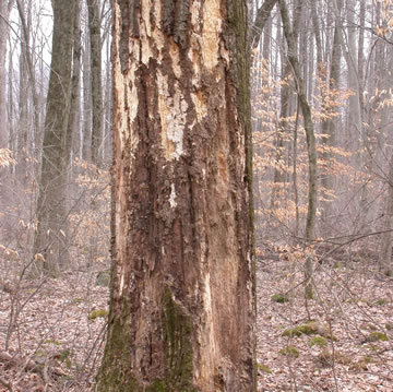
Figure 6. The same tree as in Figure 1 but observed in
March of 2014. The bark has fallen away and there was
no sign of Asian beauty. Photo © Gary Emberger.
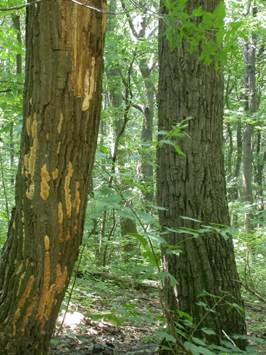
Figure
7. Asian beauty on one of two side-by-side chestnut
oak trees on June 2015 on the Darlington Trail east of Miller's
Gap Road, Cumberland County, PA. Photo © Gary Emberger.
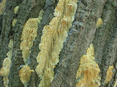
Figure 8. Spines of Asian beauty on the tree in Figure 7. On
chestnut oak trees, at least, the fungus develops in the furrows
of the bark.
Photo © Gary Emberger.
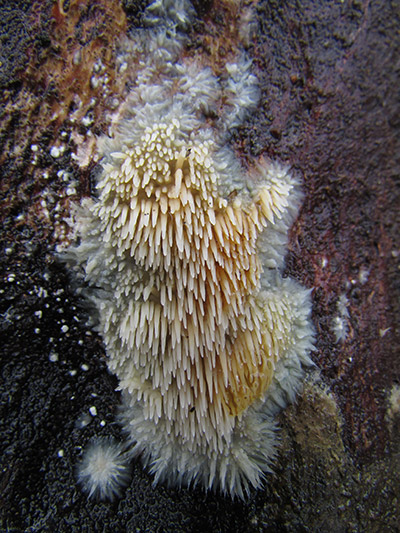
Figure 9.
This young specimen of Radulomyces copelandii
was found in Atco, NJ in an area
called the Pine Barrens.
Photo © Maricel Patino.
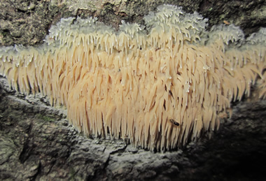
Figure 10. A very young Asian beauty fruit body developing
on the bark of
an uprooted oak tree at the Robinson Nature
Center in Howard
County, Maryland, in June of 2015.
Photo © Joanne Solem.
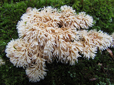
Figure 11.
Radulomyces paumanokensis - a newly reported
species
in eastern North America. The type species was
collected
from a rotted hardwood log on Long Island, New
York. The specific epithet paumanokensis is derived from
Paumanok, the Native American name for Long Island, the type
locality. The specimen above was growing in New Jersey.
It is included on this page
as a potential look-alike for
R. copelandii. Unlike R. copelandii, R. paumanokensis
fruitbodies are not as effused. They are
described as compact,
hemispherical to ovoid in shape (up to 50 x 50 mm). In
addition, the spines (up to 20 mm long) are more highly
branched. Fresh fruitbodies are white to orange-white to
pale orange, drying to pale orange, grayish-orange or
orange-gray. Photo © Maricel Patino.
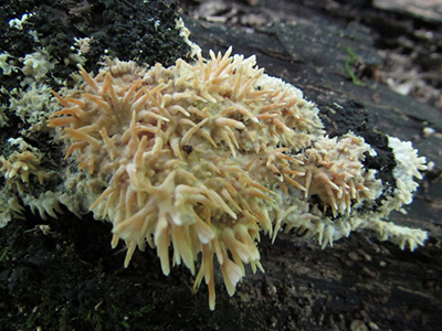
Figure
12. Another specimen of Radulomyces paumanokensis
from NJ. Photo © Maricel Patino.