Auricularia angiospermarum
Scientific name: Auricularia angiospermarum Y.C. Dai,
F. Wu, and D. W. Li
Derivation of name: Auric- refers to "ear" and the ear-like
lobes this fungus often forms. angiospermarum means
this species grows on angiosperm (flowering plant) wood.
Misapplied names: Auricularia auricula (Hooker)
Underwood; Auricularia auricula-judae (Fr.) J. Schrot.;
Hirneola auricula-judae (L.) Berk.
Common name(s): Tree-ear; Wood-ear
Phylum: Basidiomycota
Order: Auriculariales
Family: Auriculariaceae
Occurrence on wood substrate: Saprobic; hardwoods;
spring through fall.
Dimensions: 3-15 cm wide; ear-shaped to irregularly
cup-shaped, sometimes fused together.
Sterile outer surface: Usually the upper surface; yellowish-
brown
to reddish brown or grayish-brown; minutely hairy;
often ribbed or veined.
Fertile inner surface: Usually facing downward; yellowish-
brown
to reddish-brown; hairless; often ribbed or veined.
Edibility: Edible
Comments:
Auricularia auricula, the name found in many
North American field guides, is a European species that
does not occur
here. There are several North American
species with two occurring in the Northeast: Auricularia
angiospermarum grows on hardwoods (flowering plants)
and Auricularia americana grows on conifers (softwoods).
The two species differ in microscopic details
More information at MushroomExpert.com:
More information at TomVolkFungi.net
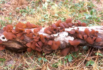
Figure
1. Wood-ears
clustered on a log.
Photo
© Larry Grand.
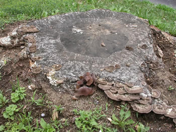
Figure 2. A cluster of tree-ears is growing to the left of
the polypore fungi in the lower right portion of this stump.
Photo © Gary Emberger.
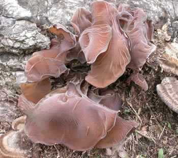
Figure 3. The cluster of tree-ears referenced in Fig. 2.
Note the wrinkling or folding of the upper, sterile surface.
Photo © Gary Emberger.
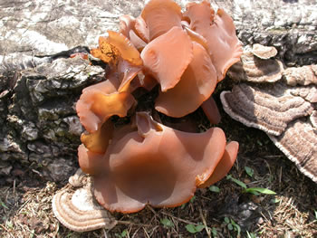
Figure 4. These are the fungi shown in Fig. 3 but after a
day of rain. Many of the wrinkles and folds have
disappeared. Photo © Gary Emberger.
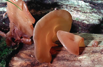
Figure 5. A typical reddish-brown specimen of tree-ear.
Photo © Pam Kaminski.
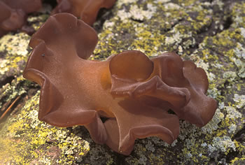
Figure 6. Note the wavy or lobed margin which is quite
typical
of wood-ears. Photo
© Fred Habegger.
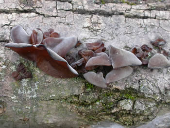
Figure 7. These specimens were found November 30,
2003,
illustrating how late in the season they can be found.
The
upper, sterile surfaces are conspicuously gray due to
the
presence of minute hairs. Photo © Gary Emberger.
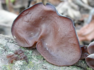
Figure 8. The fertile surface of one of the specimens
shown
in Fig. 7. Photo © Gary Emberger.
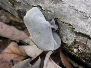
Figure 9. The finely pubescent gray sterile surface
of one of the November 30 specimens.
Photo © Gary Emberger.
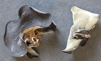
Figure 10. These dried specimens from the
collection
illustrated in Fig. 7 show the typical blackish color
observed
upon drying. The specimen on the right is gray
due to a layer
of fine hairs. Photo © Gary Emberger.