Armillaria spp.
Scientific name: Armillaria gallica Marxm. &
Romagn.; Armillaria gemina Berube & Dessur.;
Armillaria solidipes Peck; Armillaria
sinapina
Berube & Dessur.
Derivation of name: Armillaria means "with bracelets,"
referring to the ring on the stipe of many species.
Phylum: Basidiomycota
Order: Agaricales
Family: Physalacriaceae
Occurrence on wood substrate: Most species are
parasitic,
some are saprobic; depending on the species,
they may occur
as
solitary
specimens or in cespitose
clusters, they may appear
terrestrial or fruit on visible
wood, and they may be
associated with hardwood or
conifer tree species; many form
black rhizomorphs
allowing for tree-to-tree spread of the fungus.
Dimensions: Consult field guides.
Cap: Smooth or scaly, depending on the species. Consult
field
guides.
Gills: Attached.
Spore print:White.
Stipe: Consult field guides. In species with a cespitose
growth
habit, each stipe base tapers to a point.
Veil: Present (but lacking in A. tabescens).
Edibility: The various species are considered edible
which is not surprising given that they were all (other than
A. tabescens)
once considered variants of A. mellea.
Even so, some individuals will experience GI distress after
eating this species.
Comments: The websites below include information on a
number of Armillaria species found east of the Rocky
Mountains. Two of the sites include keys to species. A
number of Armillaria spp. can be identified
with certainty only through microscopic examination.
More information (key to species) at MushroomExpert.com:
More information (A. solidipes) at MushroomExpert.com:
More information (A. gallica) at MushroomExpert.com:
More information (key to species) at TomVolkFungi:
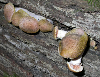
Figure 1. A. gallica. Photo © Gary Emberger.
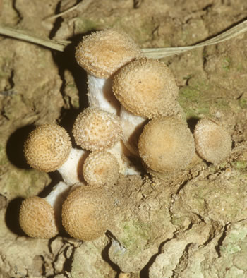
Figure 2. A. gallica. Photo © John Plischke III.
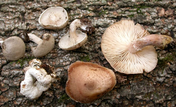
Figure 3. A. gallica and Entoloma abortivum.
Photo © Gary Emberger.

Figure 4. A. gemina. Photo © John Plischke III.
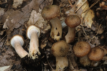
Figure 5. A. solidipes. Photo © John Plischke III.

Figure 6. A. solidipes. Photo © John Plischke III.
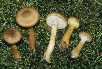
Figure 7. A. sinapina. Photo © John Plischke III.