Niveoporofomes spraguei
Scientific name: Niveoporofomes spraguei (Berk. & M.A.
Curtis) B.K. Cui, M.L. Han & Y.C. Dai
Derivation of name: Spraguei indicates this fungus species
was named after C. J. Sprague.
Synonyms: Fomitopsis spraguei (Berk. & M.A.
Curtis) Gilb. & Ryvarden; Polyporus spraguei Berk. &
M.A. Curtis; Tyromyces spraguei (Berk. & M.A. Curtis)
Murrill
Common name(s): None.
Phylum: Basidiomycota
Order: Polyporales
Family: Fomitopsidaceae
Occurrence on wood substrate: Saprobic and parasitic;
sessile to effused-reflexed on dead wood and at the bases of
living hardwoods, particularly oak; year-round.
Dimensions: Caps up to 15 cm wide.
Upper surface: Caps white to ochraceous to gray; margin
may be somewhat reddish on fresh, growing specimens;
margins of young specimens
develop a green color upon
handling;
azonate; smooth or slightly grooved, glabrous to
appressed-tomentose.
Pore surface: White to cream-colored to pale brown; pores
3-6 per mm.
Edibility: Inedible.
Comments: Young specimens exude watery drops from the
margin and upper surface. The green discoloration of the
margins
is a useful marker for this species, helping to
distinguish it from
similar species otherwise differentiated
only by microscopic
characters.
More information at MushroomExpert.com
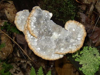
Figure 1.
The grayish cap of this young Niveoporofomes
spraguei specimen is exuding drops of water. Photo
© Cathy Cholmeley-Jones.
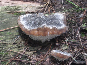
Figure 2. Water drops exuding from the pore surfaces as
well as the upper surfaces of these young specimens. Note
the pale reddish coloration at the margin. Photo © Rick
Van de Poll.
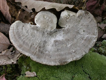
Figure 3. The cap of Niveoporofomes spraguei is
sometimes described as having the appearance of stone
or marble.
Photo © Gary
Emberger.
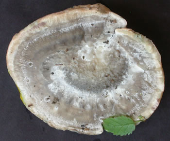
Figure 4.
Top surface of a specimen collected during a
NEMF foray. Photo © Gary Emberger.
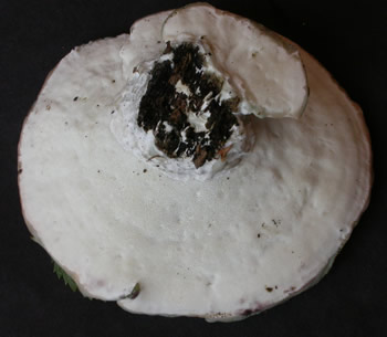
Figure 5. Bottom surface of specimen in Figure 4.
Photo © Gary Emberger.
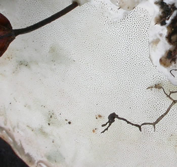
Figure 6. Pores (3-6 per mm) visible on the white pore
surface of Niveoporofomes spraguei. Photo © Gary
Emberger.
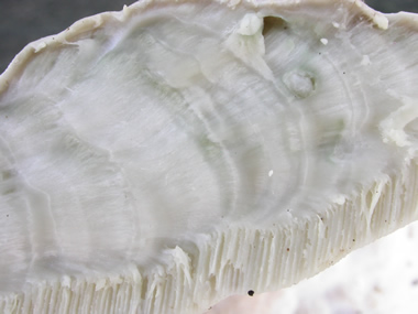
Figure 7.
This sectioned cap reveals that the thick context
(flesh)
above
the pore layer has the
same marble-like
appearence
as the cap. Photo © Gary
Emberger.
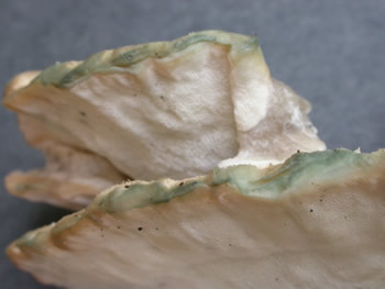
Figure 8. The margins on these fresh specimens have
discolored green where they were handled.
Photo © Gary Emberger.
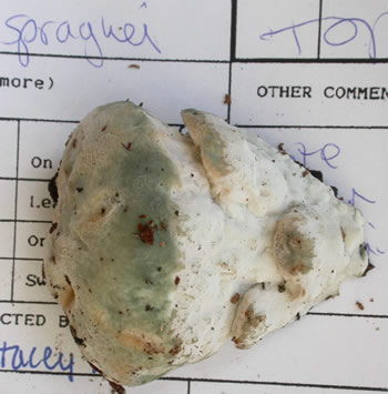
Figure 9. Young speciemen collected at a NEMF foray.
Note the green discoloration where handled.
Photo © Gary Emberger.