Apioperdon pyriforme
Scientific name: Apioperdon pyriforme (Schaeff.)
Vizzini
Derivation of name: Pyri- means "pear" and form-
means
"shape" or "appearance." Pyriforme refers to the
pear shape
of this puffball.
Synonyms: Lycoperdon pyriforme Schaeffer:Persoon;
Morganella pyriformis (Schaeffer:Persoon)
Kreisel & D. Kruger
Common name(s): Pear-shaped puffball.
Phylum: Basidiomycota
Order: Agaricales
Family: Agaricaceae
Occurrence on wood substrate: Saprobic;
scattered or in
dense clusters on decaying wood; July through November.
Dimensions: Fruit bodies are 1.5-4.5 cm wide and 2-5 cm
tall.
Description:This puffball species is pear-shaped to nearly
globose and supported by a small sterile base attached to the
substrate by white mycelial strands (rhizomorphs). When
young this puffball
is whitish and covered with tiny warts and
granules. With maturity the spore case (peridium) is yellow-
brown to reddish-brown and develops a pore-like
mouth (the
ostiole) at the apex allowing
spores to be "puffed out" when
the outer case is disturbed by
raindrops or twigs striking it.
The spore
producing internal tissue (gleba) is moist and white
at first,
turning olive-brown and powdery when mature.
Edibility: Edible.
Comments: This species is edible only when the internal spore
tissue (gleba) is completely white and uniform in appearance.
Care must be taken not to confuse puffballs with young stages
of Amanita species which are enclosed by a universal veil.
A longitudinal section of a young Amanita will reveal some
tissue differentiation into gills. Gills never occur in puffballs.
More information at MushroomExpert.com:
More information at TomVolkFungi.net:
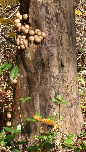
Figure 1. Clustered specimens of Apioperdon pyriforme
on a standing dead tree.
Photo © Gary Emberger.
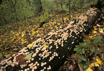
Figure 2. Dense clusters of Apioperdon pyriforme on a
rotting log. Photo
© Fred Habegger.
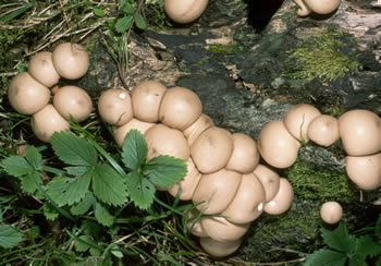
Figure 3. A closer view of young specimens of
Apioperdon pyriforme. Photo © William Roody.
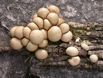
Figure 4. This detached piece of bark bearing a cluster of
pear-shaped puffballs was brought to the display tables at
a NEMF foray. The bark was placed on a stone wall for
the photograph. Photo © Gary
Emberger.
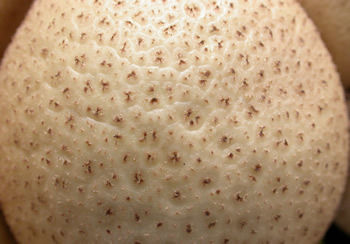
Figure 5. A close-up of the surface of one of the young
puffballs
in Figure 4. Field guides describe the surface of
young specimens
as
covered with minute warts, particles,
or granules. Compare this to young specimens of
Lycoperdon perlatum which are covered
with conical
spines.
Photo © Gary Emberger.
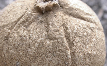
Figure 6. The peridium of a mature specimen. An ostiole
has developed at the top of the puffball. Photo © Gary
Emberger.
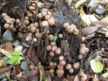
Figure 7. Puffballs growing on a stump. Ostioles are
developing on many of these. Photo © Gary Emberger.
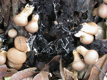
Figure 8. Dislodging some of the puffballs on the stump
in Figure 7 reveals white rhizomorphs extending from the
puffball bases
to the woody substrate, a useful
field
identification character. Photo © Gary Emberger.
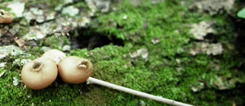
Figure 9. These mature speciemens have developed
pores for release of the spores. The small stick was
used to gently nudge the puffball near it. See Figure 10
for the result.
Photo © Gary Emberger.
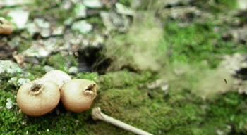
Figure 10.
These are the puffballs of Figure 9, a moment
after the stick was used to compress the puffball. A
cloud of brownish spores was released - visible on the
right
side of the photograph.
Photo © Gary Emberger.
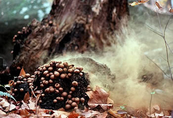
Figure 11.
Can you see this spore cloud?!
Photo © Pam Kaminski.
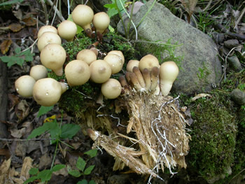
Figure 12.
White rhizomorphs of Apioperdon
pyriforme are evident
throughout the piece of well-rotted
wood broken away from a log bearing a number of
very
immature
puffballs. The young puffballs are highlighted
in
Figure 13. Photo © Gary Emberger.
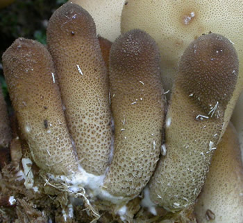
Figure 13.
Close-up of the tall, cylindrical immature
specimens of
pear-shaped puffball visible in Figure 12. It
was interesting to see the puffballs grow almost to mature
height
before the fertile portion expanded - perhaps
reflective of the crowded growing conditions.
Photo © Gary Emberger.