Scleroderma citrinum
Scientific name: Scleroderma citrinum Persoon
Derivation of name: Sclero- means "hard" and derma
refers to "skin." Citrin- means "lemon-yellow."
Synonyms: S. aurantium (L.) Pers.; S. vulgare Hornem.
Common name(s): Common earthball; Golden
Scleroderma; Pigskin poison puffball.
Phylum: Basidiomycota
Order: Boletales
Family: Sclerodermataceae
Occurrence on wood substrate: Mycorrhizal; solitary or
grouped on the ground or on decaying wood; July through
November.
Dimensions: Fruit bodies are 2.5-10 cm wide and 2-4 cm
tall.
Description: The nearly globose to somewhat flattened fruit
bodies are pale brown to yellow-brown and covered with
large, coarse, flattened warts. At maturity they open
irregularly at the top to expose the spores. When young the
internal flesh (gleba) is firm and white but soon becomes
marbled and purplish-black to black and firm until at
maturity, the spore mass is powdery and blackish-brown.
Sectioning reveals the thick rind-like white outer wall
(peridium) which may be up to 4 mm or more thick.
Edibility: Poisonous.
Comments: Read about the relatedness between this
"puffball" species and boletes at the website below.A
look-alike species to Scleroderma citrinum is
Scleroderma areolatum but S. areolatum generally
has a thinner peridium and is covered by minute, dark
brown scales. Scleroderma citrinum is sometimes
parasitized by the bolete, Pseudoboletus parasiticus
(see Figure 10).
More information at MushroomExpert.com:
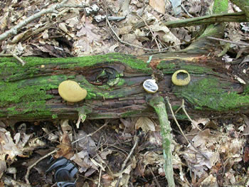
Figure 1.
Scleroderma citrinum on a rotting log. The
specimen in the middle was cut in half. The sectioned
specimen and the older specimen on the far right are
in closer view in Figure 8. Photo © Gary Emberger.
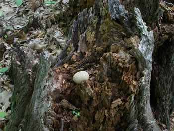
Figure 2. Scleroderma citrinum fruiting on the top of a
rotting stump. Photo © Gary Emberger.
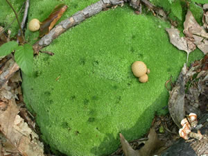
Figure 3. Pigskin poison puffball does not always grow
on wood. There was no wood under this specimen
growing in a bed of moss.
Photo © Gary Emberger.
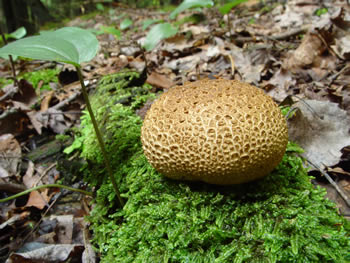
Figure 4. A typical specimen of Scleroderma citrinum.
Note the rough pattern of warts on the surface. Photo ©
David Work.
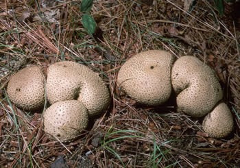
Figure 5. The common earthball
is very firm to the touch
and never has the feel of the true puffballs.
Photo © William
Roody.
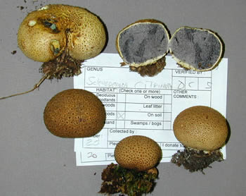
Figure 6. A variety of specimens collected during a NEMF
foray. A mass of mycelial strands connect the fungus to the
substrate. Particles of substrate held by these strands are
visible on the specimens at the bottom of the picture.
Photo © Gary Emberger.
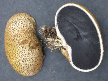
Figure 7.
A sectioned specimen reveals the thick white peridial
wall and the firm, dark purplish-black glebal mass.
Photo © Gary Emberger.
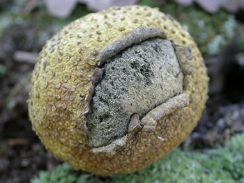
Figure 8. Unlike Lycoperdon species,
Scleroderma
citrinum does not form a well-defined ostiole. Instead,
the peridium ruptures to form an irregular opening through
which the spores are released. Photo © Gary Emberger.
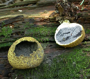
Figure 9. The mature specimen on the left is has opened via a
ragged apical pore. The younger, longitudinally-sectioned
specimen on the right shows the marbled flesh with white
veining characteristic of the species. Photo © Gary Emberger.
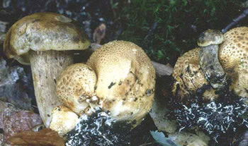
Figure 10. Scleroderma citrinum
parasitized by the bolete,
Pseudoboletus parasiticus. Photo © George Barron.