Mutinus elegans
Scientific name: Mutinus elegans (Mont.) E. Fisch.
Derivation of name: Mutinus was a Roman phallic
deity and
means "penis," elegans means "elegant."
Synonyms: Corynites elegans Mont.
Common name(s): Elegant stinkhorn,
Devil's dipstick.
Phylum: Basidiomycota
Order: Phallales
Family: Phallaceae
Occurrence on wood substrate: Saprobic; solitary to
grouped on soil, mulch, wood chips, and decaying wood;
June through September.
Dimensions: Fruitbody 10-18 cm tall and 1.5-2.5 cm thick.
Description: Fruit body at first a white to pinkish egg-like
stage, resembling a puffball. The "egg" is attached to the
substrate by white mycelial strands (rhizomorphs). The
outer wall (peridium) of the "egg" splits and a hollow,
spongy, stalk extends outward. The bumpy, minutely
pitted and typically curved stalk may be whitish below and
orangish to orange-red to reddish above or uniformly
colored.
The stalk tapers gracefully from about the middle
to the tip and may be slightly curved. A slimy, olive-brown,
fetid spore mass covers the upper
1/3 (up to 6 cm) or more
of the fruit body. This spore zone is not clearly marked-off
from the rest of the stalk.
Edibility: Said to be edible in the egg stage.
Comments:
Flies are attracted to the fetid slimy mass and
serve to disperse the spores. This species and two closely
related species - M. caninus and M. ravenelii may be
difficult to cleanly separate from
each other. Consult
the website below for additional
comments on these
three species.
More information at MushroomExpert.com:
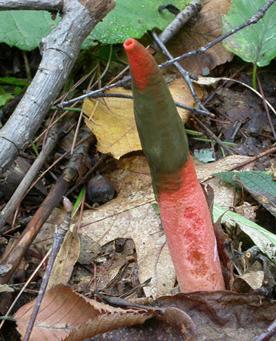
Figure 1. Mutinus elegans growing in a woodland setting.
Photo © Gary Emberger.
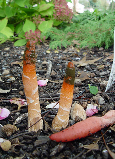
Figure 2. The upright specimens are much more orangish
in coloration than the specimen in Figure 1. In general,
the intensity of coloration in Mutinus species often
diminished towards the base of the
stalk.
Photo
© Geroge Weigel.
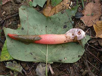
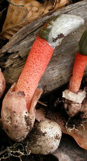
Figure 3. The gracefully tapering stalk and a spore mass
covering at least 1/3 of the stalk are good field characters
for Mutinus elegans. Note the
rhizomorphs attached to
the base of the egg.
Photo © Gary Emberger.
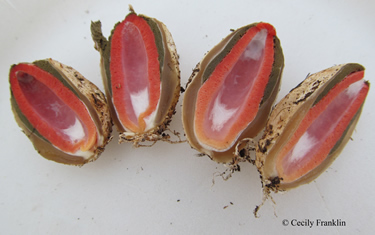
Figure 4. Dissected eggs of Mutinus elegans.
The peridium,
dark spore mass on the upper stalk, and the reddish stalk are
all
clearly observable.
Photo © Cecily Franklin.
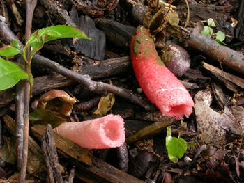
Figure 5. The stalks of Mutinus species are hollow.
Photo © Gary Emberger.
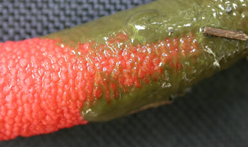
Figure 6. The
reddish stalk can be
seen
where some of
the greenish spore mass was rubbed
away. In M.
elegans, the slimy
spore-bearing
zone is not usually
clearly marked-off from the rest of the stalk
Photo © Gary Emberger.
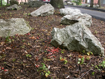
Figure 7.
A dense cluster of Mutinus elegans growing in
a heavily wood-mulched landscape bed.
Photo © Gary Emberger.
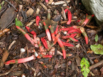
Figure 8. Close up of the cluster in Figure 7. Some of
the specimens have a strikingly reddish coloration.
Photo © Gary Emberger.
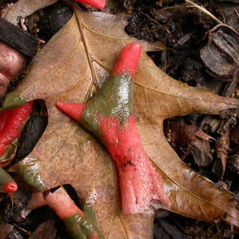
Figure 9.
This specimen has an interesting forked,
spore-bearing portion.
Photo © Gary Emberger.
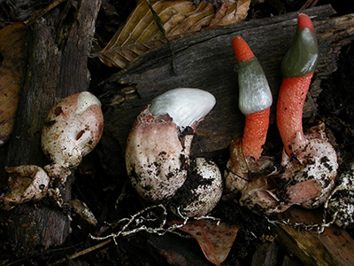
Figure 10. The elongating stalks of the two specimens to the
left are visible as they break
through the egg-like peridium.
This photograph was taken at 9:00 AM. See Figure 11.
Photo © Gary Emberger.

Figure 11. The same specimens shown in Figure 10 but
photographed 12 hours later. The stalk of the second specimen
from the left
has elongated
considerably.
Photo © Gary Emberger.
Figure
12. The stalk is noticably pitted in some areas
but perhaps not as conspicuously as occurs in
M. ravenelii. Depending on the specimen, M. elegans,
M. caninus and M. ravenelii may be
difficult to clearly
differentiate from
each other.
Photo © Gary Emberger.