Pseudocolus fusiformis
Scientific name: Pseudocolus fusiformis (E. Fisch.)
Lloyd
Derivation of name: Fus- means "spindle," form-
means
"shape or appearance" in reference to the long,
spindle-shaped arms.
Synonyms: Colus fusiformis E. Fisch.; Pseudocolus
schellenbergiae (Sumst.) P. Micheli
Common name(s): Stinky squid.
Phylum: Basidiomycota
Order: Phallales
Family: Phallaceae
Occurrence on wood substrate: Saprobic; single to
several in wood chips used for landscaping, wood debris
or leaf litter;
July through September.
Dimensions: Fruitbody up to 6 cm tall and 1.5 to 3 cm
wide..
Description: Fruitbody at first a brownish to grayish
egg-like
stage, resembling a puffball, with white
rhizomorphs. When the outer wall (peridium) of the egg
splits open, three to five slender, tapering, pink to orange
arching arms rise from a common stalk. The arms are
whitish at their bases and the tips are often united. The
greenish, slimy, fetid spore mass covers the inner
surfaces of the arms.
Edibility: Inedible.
Comments: Gary Lincoff reports that this species was
first reported in North America in Pittsburgh in 1915
and has spread widely since then.
More information at MushroomExpert.com:
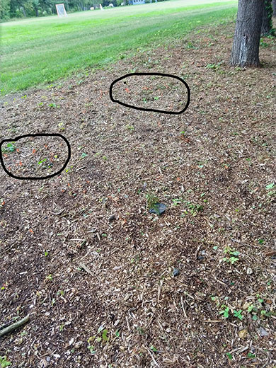
Figure 1. The photographer smelled the specimens of
Pseudocolus fusiformis before she
saw them growing in this
wood-mulched area of a park. The pinkish-orange specimens are
barely visible in the circled areas. Photo
© Melissa Emberger.
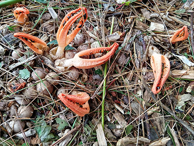
Figure 2. A closer view of the stinky squid specimens in Figure 1.
In addition to the colorful fruitbodies, note the cluster of
brownish-gray eggs from which they emerge.
Photo
© Melissa Emberger.
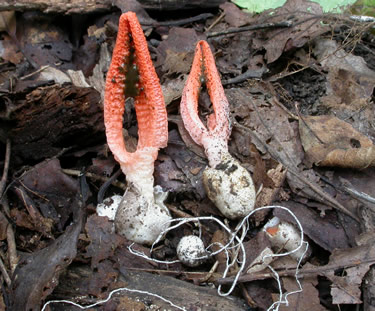
Figure 3. Two
unopened
"eggs" are at the bottom right. Both
they
and the mature specimen on the right were
detached from
the substrate in order to show the white rhizomorphs.
Photo © Gary Emberger.
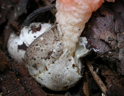
Figure 4. Stalk emerging from ruptured "egg" of
Pseudocolus
fusiformis. Photo © Gary Emberger.
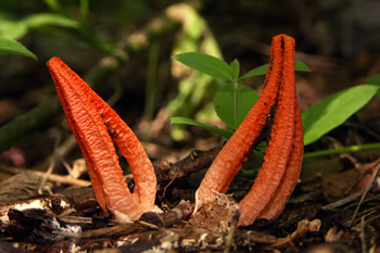
Figure 5. This young specimen shows the dark spore mass
surrounded by the "arms." Photo © Fred Habegger.
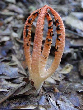
Figure 6. The tips of the arms are often held together and
the spore mass is distributed along the inner surfaces of the
arms. Photo © Dorothy Smullen.
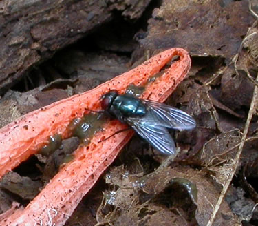
Figure 7. As always, flies find the spore mass of stinkhorns
irresistible.
Photo © Gary Emberger.